top of page



Before you start
Select your target protein (or proteins)
Select your loading controls (we recommend GAPDH and/or Vinculin, but others are acceptable)
Select your membrane (PVDF or Nitrocellulose) for benchmarking, a side-by-side experiment is the best way to see improvements in your protocol.
Plan for dilutions (e.g: 20, 10, 5, 2.5, 1.2, 0.6, 0.3, 0.15, μg). We have detected target protein with loading as little as 0.15 μg of lysate. (this will depend in quality of Ab)
Running the WB
Activate your membrane (no methanol needed)
15 s in EtOH >> 2 min Transfer Buffer >> Ready to GO
Wet Transfer (30-90 V, 60-120 min)
Optimize based on your desired protein MW. For sharp bands always go for low voltages and higher times (Start with 45 V, 2h).
Semi-dry transfer (0.4 A constant, 25 V, 20-30 min)
Follow the recommended settings to ensure good protein transfer without errors. See better results with chilled buffer.
If you are using Turbo conditions (< 10 min) with BioRad Trans-Blot Turbo buffer then please use 0.8 A (constant), 20 V and 7-10 min. See better results with chilled buffer.
See the results
Block the membrane with your usual blocking buffer
Add primary antibody at your usual dilution
Add secondary antibody at your usual dilution
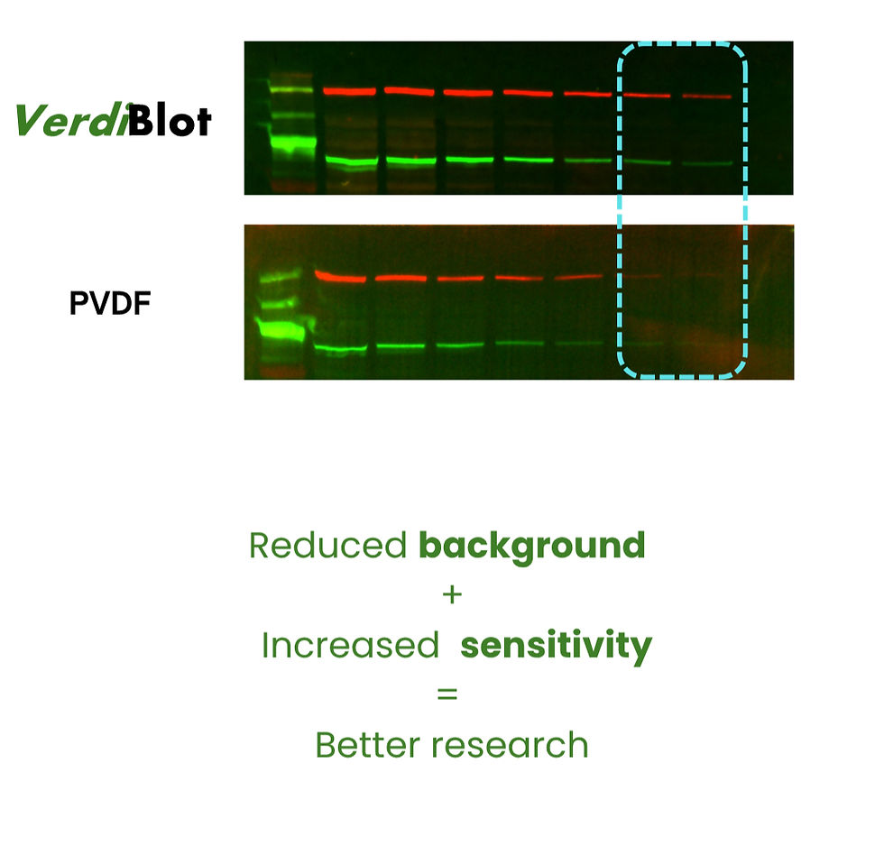
Compare the membranes!
- Qualitative test- Are all the target bands there?
- Quantify the band intensity- Use your favorite imaging software to see the real band intensity
- Background- compare the background between membranes, specially at long exposure times.
Stripping and Reprobing (Optional)
VerdiBlot is suitable for stripping and reprobing. Optimization is always recommended but this guide walks you through the main process.
Simplified protocol
Start with mild conditions. Submerge the membrane in stripping buffer (S. buffer: 0.1 M glycine, pH 2.2, 0.1% SDS, 1% Tween-20) and agitate for 10-20 min. Rinse the membrane with TBST/PBST buffer for 10 min (3X). Before continuing, add ECL substrate and develop the blot, you should see no signal, this indicates the initial Abs were successfully stripped away. If signal is present, repeat step 1. Block the membrane and proceed to reprobe with primary and secondary antibody as you did the first time (different primary Ab). For more details see the Full Guide here.
Compatibility of the membrane
Detection method
Fully compatible with Chemiluminescence and Fluorescence detection.
Fully compatible with monoclonal and polyclonal antibodies
Blocking agents
Fully compatible with common blocking agents:
Casein, dry milk, BSA, synthetic and commercial brands (e.g: Azure Blot Blocking buffers)
Total Proteins Stains
Ponceau S, Coomassie Brilliant Blue, colloidal gold, and more.
See other Results

Frequently asked questions
FAQ EN
Add a Title
bottom of page